Articular cartilage separates the bones in articulating joints (e.g. hips, knees, shoulder, etc.) and allows for almost effortless movement. The predominant cell types found in articular cartilage are termed chondrocytes and are responsible for building, maintaining and degrading the tissue. The pathological breakdown of articular cartilage results in osteoarthritis, which is a wide-spread disease in western civilizations and has a major socio-economic impact. An active lifestyle and adequate mechanical stimulation are essential for cellular health and tissue maintenance. However, to date the molecular mechanisms on how chondrocytes (cartilage cells) integrate mechanical forces into a cellular response (mechanotransduction) are not fully understood. Our aim is, therefore, to better understand the effects of mechanotransduction on cartilage degeneration and regeneration.
Among other mechanisms, mechanosensitive ion channels are thought to play a key role in mechanotransduction. Ion channels are a major contributor to the cell’s electrical potential across the cell membrane (membrane potential). Previous experiments have shown that the membrane potential is gravity dependent in neurons and cartilage cells. In this experiment, we study alterations in membrane potential and, in particular, the concentration of free calcium in chondrocytes in response to altered gravitational conditions during parabolic flights. Calcium is a common signaling molecule for a number of cellular processes and is thought to enter the cell through mechanosensitive ion channels.
We used two techniques to measure the cellular free calcium. On the one hand, an engineered protein is introduced in the cells. This fluorescent protein undergoes an irreversible change in the presence of calcium and UV light. This subsequently results in a change in fluorescent color. Therefore, by the fluorescent colors, the concentration of cytosolic free calcium, at the moment where the cells were briefly irradiated with UV light can be determined. On the other hand, a commercial plate reader and calcium sensitive dye was used. The cells were stained preflight with a specialized fluorescent dye, which indicates changes in cytosolic calcium concentration. This fluorescent signal was then recorded during the flight. In a similar way, the membrane potential was recorded by using a voltage sensitive dye.
Chondrocytes build and maintain articular cartilage in joints. However, if this tissue is affected by a pathological breakdown, osteoarthritis results, which is a widespread disease in western civilizations. Our aim is to better understand the mechanotransduction pathways of chondrocytes. The results may one day impact our understanding and treatment of osteoarthritis.
75th ESA Parabolic Flight
73rd ESA Parabolic Flight
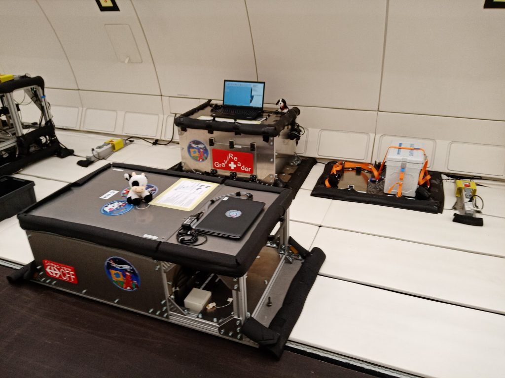
Rack in the aircraft 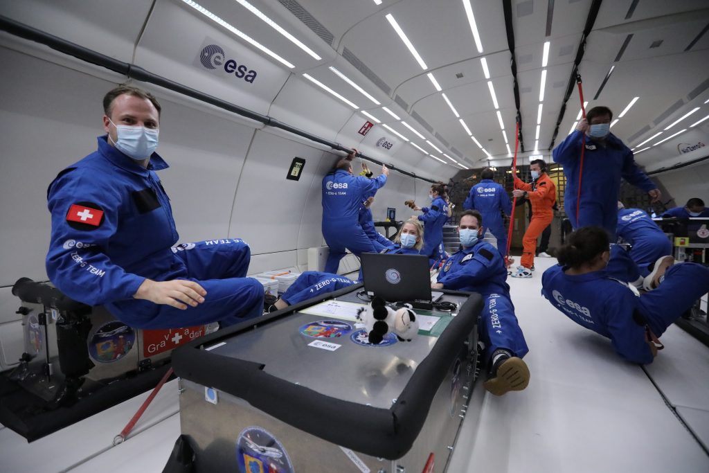
Zero Gravity 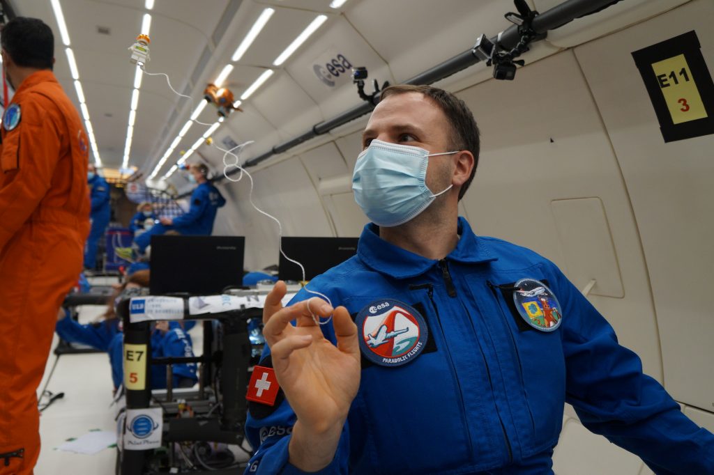
Legonaut